Skin Cancer Examination
Skin cancer is one of the most common and most treatable cancers.

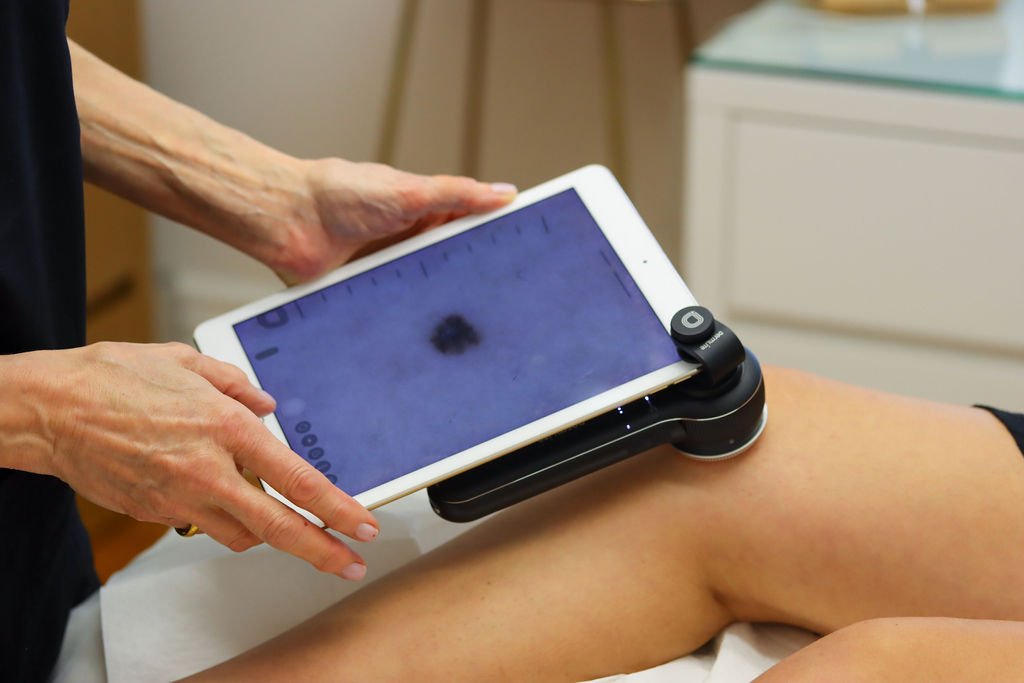
I recommend and regularly perform head to toe skin cancer examinations for patients. Some patients are more at risk than others, with risk largely depending on the degree and type of sun exposure, as well as skin type. During a Skin Cancer Examination a history is taken to determine risk factors and to help determine the skin type. Every skin lesion is then examined using a dermascope (skin microscope which enlarges the lesion 10 X) and all suspicious lesions are photographed through the dermascope and these images are recorded on the patient’s file.
Skin cancers are divided into two categories- Melanoma and Non Melanoma Skin Cancer (NMSC). NMSC is further divided into two categories – Squamous cell carcinomas (SCC) and Basal cell carcinomas (BCC). If a lesion is suspicious for melanoma, it is completely excised and sent to the laboratory for tissue diagnosis.
New Zealand has the highest incidence of melanoma in the world.

Certain NMSCs (SCC) that arise on the face and scalp can metastasize (spread throughout the body) in much the same way a melanoma behaves and so it is important to make the correct diagnosis for NMSCs. Sometimes the diagnosis is not obvious using the dermascope and a punch biopsy is necessary to make a tissue diagnosis.
A punch biopsy utilises a disposable circular blade to collect a sample of tissue from the suspicious skin lesion. First local anaesthetic is injected around the lesion so the procedure is painless. The punch biopsy blade is then inserted into the centre of the lesion to produce a circular tube of tissue containing a sample of skin from the top layer down to the fatty layer below. This punch biopsy specimen is put in a pot containing preservative solution and sent to the laboratory.
A photographic record of your moles is critical.
If a skin lesion is suspicious I will explain why that is so using the photographic images taken through the dermascope. If a punch biopsy or skin cancer excision surgery is required I can either perform the procedure or refer to a plastic surgeon if required.
Photographic images of moles that are slightly abnormal are stored securely on the patient’s file and follow-up examination and photography is recommended in order to determine if the mole is changing. If it is changing, it should be removed.
